


 |
| Animated Form of Human Skeleton |
 |
| Human Skeleton System |
Human Skeleton System:
The human skeleton consists of both fused and individual bones supported and supplemented by ligaments, tendons, muscles and cartilage. It serves as a scaffold which supports organs, anchors muscles, and protects organs such as the brain, lungs and heart.
The biggest bone in the body is the bone in the thigh and the smallest is the stapes bone in the middle ear. Several factors contribute to the bone density and average mass of the human skeleton including; gender, race, hormonal factors, nutrition, physical activity and lifestyle behaviors. Because of these and other factors affecting an individual's weight the human skeleton may comprise between 12 and 20 percent of a person's total body weight with the average being 15 percent.
Fused bones include those of the pelvis and the cranium. Not all bones are interconnected directly: there are three bones in each middle ear called the ossicles that articulate only with each other. The hyoid bone, which is located in the neck and serves as the point of attachment for the tongue, does not articulate with any other bones in the body, being supported by muscles and ligaments.
At birth, a newborn baby has over 300 bones, whereas on average an adult human has 206 bones (these numbers can vary slightly from individual to individual).
 |
| Human Hand and Collar Bone |
Collar Bone:
CLAVICLE(COLLAR BONE):
The clavicle is an S-shaped long bone.It articulates with the manubrium of the sternum at the sterno-clavicular joint and forms the acromioclavicular joint with the acromion process of the sacpula.The clavicle provides the only bony link between the upper limb and the axial skeleton.
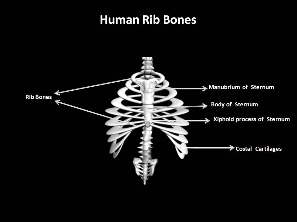 |
| Human Chest Bone |
Chest Bones:
STERNUM(BREAST BONE):
The flat bone can be felt just under the skin in the middle of the front of the chest. The manubrium is the upper most section and articulates with the clavicles at sternoclavicular joints and with the first 2 pairs of ribs. the body of middle portion gives attachment to the ribs.The xiphoid process is the tip of bone.It gives attachment to the diaphragm, muscles of the anterior abdominal wall and the linea alba.
 |
| Human Hip Bone |
Hip Bones:
PELVIS:
The pelvis is formed by the hip bones, the sacrum and the coccyx.It is divided into upper and lower parts by the brim of pelvis,consisting of the promotory of the sacrum and the iliopectineal lines of the innominate bones.The greater or false pelvis is above the brim and the lesser or true pelvis is below.
The shape of the female pelvis allows for the passage of the baby during childbirth. In comparison with the male pelvis, the female has lighter bones,is more shallow and rounded and roomier.
 |
| Human Skull Bone |
Human Skull Bones:
CRANIUM:
The cranium is formed by a number of flat and irregular bones that provide a bony protection for the brain. It
has a base upon which the brain rests and a vault that surround and cover it.In mature skull the joint (sutures) between the bones are immovable(fibrous). the bones have numerous perforation(foramina,fissures) through which nerve blood and lymph vessels pass.The bones cranium are:
1 frontal bones
2 parietal bones
2 temporal bones
1 occipital bone
1 sphenoid bone
1 ethmoid bone.
ETHMOID BONE:
This bone occupies the anterior part of the base of the skull and helps to form the orbital cavity,the nasal
septum and the lateral wall of the nasal cavity.On each side are 2 projections into the nasal cavity The upper
and middle conchae or tubinated processes.It is a very delicate bone containing many air sinuses lined with
ciliated epithelium and with openings into the nasal cavity.The horizontal flattened part,the cribriform
plate,forms the roof of the nasal cavity and has numerous small foramina through which nerve fibres of
the olfactory nerve pass upward from the nasal cavity to the brain.There is also a very fine perpendicular
plate of the bone that forms the upper part of the nasal septum.
MANDIBLE:
This is the lower jaw,the only movable bone of the skull.It originates as 2 parts that unite at the midline.Each half consists of 2 main parts: a curved body with the alveolar ridge containing the lower teeth and a ramus, which projects upwards almost at right angles to the posterior end of the body.
MAXILLA(UPPER JAW BONE)
This originates as 2 bones,but fusion takes before the birth.The maxilla forms the upper jaw,the anterior part
of the roof of the mouth,the lateral wall of the nasal cavity and part of the floor of the orbital cavities.
The alveolar ridge or process,projects downwards and carries the upper teeth.On each side the large air
sinus.
OCCIPITAL BONE:
The bone forms the back of the head and part of the base of the skull.It has immovable fibrous joints with the
parietal,temporal and sphenoid bones.Its inner surface is deeply concave and the concavity is occupied by the occipital lobes of the cerebrum and by the cerebellum.The occiput has 2 articular condyles that form condyloid joints wuth the first bone of the vertebral column the atlas.This joint permits nodding movements of the head.Between the condyles is the foramen magnum('Large hole')through which the spinal cord passes into the cranial cavity.
PARIETAL BONE:
These bones form the sides and roof of the skull.They articulate with each other at the sagittal suture,with the frontal bone at the coronal suture, with the occipital bone at the lamboidal suture and with the temporal bone at the squamous sutures.The inner surfaceis concave and is grooved by the brain and blood vessels.
TEMPORAL BONE:
These bones lies one on each side of the head and form fibrous immovable joints with the parietal,occipital,
sphenoid and zygomatic bones.Each temporal bone has several important features.
The squamous part is the thin fan shaped area that articulate with parietal bone.
The zygomatic process articulate with zygomatic bones to form the zygomatic arch(cheekbone).
The mastoid bone contains the mastoid process,a thickened region behind the ear.
The petrous portion form part of the base of the skull and contains the organs of hearing(The spiral organ) and balance.
The temporal bone articulates with the mandible at the temporomandibular joint.
VOMER:
The vomer is a thin falt bone that extends upwards from the middle of the hard palate to form most of the
inferior part of the nasal septum.Superiorly it articulates with the perpendicular plate of the ethmoid
bone.
ZYGOMATIC(cheek)BONES:
The zygomatic bones form the prominences of the cheeksand part of the floor and lateral walls of the orbital
cavities.
PALATINE BONES:
These are 2 small l-shaped bones.The horizontal parts unite to form the posterior part of the hard palate and the perpendicular parts project upwards to form part of the lateral walls of the nasal cavity.At the upper extremities they form part of the orbital cavity.
 |
| Human Vertebral Column |
Back Bones:
VERTEBRAL COLUMN:
There are 26 bones in the vertebral column. 24 separate vertebrae extend downwards from the occipital bone of the skull;then there is the sacrum,formed from 5 fused vertebrae, and lastly the coccyx, or tail,which is formed from between 3 to 5 small fused vertebrae. The vertebral column is divided into different regions.The
first 7 vertebra is neck form the cervical spine;Next 12 vertebrae are the thoracic spine,and next 5 are lumber spine.The lowest vertebra of which articulates with sacrum.
CERVICAL VERTEBRAE:
1st cervical atlas below the atlas is the axis.The 7th is vertebra prominence.
THORACIC VERTEBRAE:
Larger than cervical vertebrae.
LUMBER VERTEBRAE:
These are the largest of vertebrae because they have to support the weight of the upper body.
SACRUM:
This consists of 5 rudimentary vertebrae fused to form a triangular or wedge-shaped bone with the concave anterior surface.
COCCYX:
This consist of 4 terminal vertebrae fused to form a very small triangular bone,the board base of which articulates with the tip of sacrum.
|
| Human Hand Bone |
Hand Bones:
HUMERUS:
This is the bone of upper arm.The head sits within the glenoid cavity of the sacpula,forming the shoulder
joint.Distal to the head are two roughened projectionsof bone, the greater and lesser tubercles and between
them there is a deep groove or intertubercular sulcus,occupied by one of the tendons of the biceps muscle.The distal end of the bone presents two surfaces that arti-culate with the radius and ulna to form the elbow joint.
ULNA & RADIUS:
These are 2 bones of forearm.The ulna is longer than and medial to the radius and when the arm is in the anat-tomical position, i.e. with the palm of the hand facing forward, the 2 bones are parallel.They articulate with the humerus at the elbow joint,the carpal bones at the wrist joint and with each other at the proximal and distal radioulnar joints.In addition an interosseous membrane, a fibrous joints,connects the bones along
 |
| Human Palm and Feet Bone |
CARPAL:
There are 8 carpal bones arranged in 2 rows of 4.From outside inwards they are:
Proximal Row: sacphoid, lunate, triquetral, pisiform
Distal Row : trapezium,trapezoid,capitate, hamate.
These bones are closely fitted together and held in a position by ligaments that allow a limited amount of movement between them.
METACARPAL BONES:
These 5 bones form the palm of the hand.They are number-ed from the thumb side inwards.The proximal ends
articulate with the carpal bones and the distal ends with phalanges.
PHALANGES:
There are 14 phalanges, 3 in each finger and 2 in the thumb.They articulate with the metacarpal bones and with each other, by hinge joints.
 |
| Human Leg Bone |
Leg Bones:
FEMUR:
Femur is the longest and heaviest bone of the body. Its head is almost spherical and fits in the acetabulum of the hip bone to form hip joint, the neck extend outward and slightly downward from the head of the shaft and most of it within the capsule of the hip joint
TIBIA:
The tibia is the medial of the 2 bones of the lower leg.the proximal extremity is board and flat and presents
2 condyles for articulation with the femur at the knee joint.The head of fibula articulate with inferior aspect
of lateral condyle,forming the proximal tibiofibularjoint.The distal extremity of tibia forms the ankle joint with the talus and fibula.
FIBULA:
The fibula is the long slender lateral bone in the leg.The head of upper extremity articulates with the lateral
codyle of the tibia,forming the proximal tibiofibular joint,and the lower extremity articulates with the
tibia,and project beyond it to form the lateral malleolus.This helps to stabilise the ankle joint.
PATTELA:
This is a roughly triangular-shaped sesamoid bone associated with the knee joint.Its posterior surface
articulates with the pattelar surface of the femurin the knee joint and its anterior surface is in the
patellar tendon,i.e. the tendon of the quadriceps femoris muscle.
|
| Human Palm and Feet Bone |
TARSAL(ANKLE)BONES:
The 7 tarsal bones forming the posterior part of the foot are the talus, calcaneous, navicular,cuboid and 3 cuneiform bones.The talus articulates with the tibia and fibula at the ankle joint.The calcaneous forms the heel of the foot.The other bones articulate with each other and with the metatarsal bones.
METATARSAL:
These are 5 bones numbered from inside out,which form the greater part of dorsal of the foot.At their proximal ends they articulate with the tarsal bones and at their distal ends, with the phalanges.The enlarged distal head of the 1st metatarsal bone forms the ball of the foot.
PHALANGES(TOE BONES):
There are 14 phalanges arranged in a similar manner to those in the fingers,i.e. 2 in the great toe and 3 in the each of the other toes.
3D animation showing and describing different Human Bone System.
 Nature Magazine
 National Geographic
 Cartoon Games
 History Channel
 Online Movies
 Scientific American
 Filmfare Mag
 Online Britannica
 Time Mag
 Unexplained Mysteries


Nice images, though the proportions look kinda funky.
ReplyDeleteThe picture labeled "Anatomy of Human Hand" is actually of the human arm and doesn't show the hand, which is everything distal of the radius/ulna- the hand is actually what you call the palm later on. I guess you did it this way so that both radius and ulna and their various parts are clearly visible while image is short enough to fit into a certain area, but the radius and ulna look way too far apart and also too thick compared to the humerus.
The rib cage doesn't look at all right for a human either. The ribs should be round not flat. The cranial ribs aren't that small. I think you should put the costal cartilage in a different color so that people can see the difference between cartilage and bone (if the same color was used for cartilage disks in the spine and cartilage in the joints in other figures, that would be even better, but I think this is the only image where the lack of color-coded cartilage is confusing).
I think that the pelvis is usually just called the pelvis, even by uneducated people, at least in American English. Some people might call it the hip bone sometimes, but I'm pretty sure that no one (or at least no one speaking American English) would ever call it the waist bone. If I heard someone say "waist bone" I would never figure out that they meant the pelvis but would think they didn't know what they were talking about.
I'm not sure what's going on between the eyes in the skull image- it doesn't look like that on any real skull (or mold of a skull) I've seen. The teeth look wrong; I think they should be going further back. Also, there should be 32 of them, unless it's supposed to be a child in which case there should be 20. I think teeth are kind of important to get right, because they give you information about the diet of the animal, and they are the hardest part of the body, so they are most often preserved; I've heard there have been entire extinct species proposed based on the existence of fossilized teeth without much other surviving remains.
That is incredibly annoying, the way that the ad software inserted a link into my comment (in the words "about the diet") that isn't related to what I wrote. It makes me not want to comment again.
ReplyDeleteMy site www.ptrscience.com
ReplyDelete