


 |
| Male Reproductive Organs in Animated Form |
 |
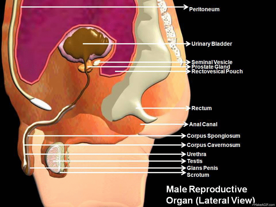
| All Male Reproductive Organs in a Single Picture |
REPRODUCTIVE SYSTEM:
The ability to reproduce is one if the properties distinguishing living from non-living matter.The mo0re primitive the animal,simpler the process of reproduction.In human being the process is one of sexual reproduction,in which the male and female organs differ anatomically and physiologically.
Both males and females produce specialized reproductive germ cell, called gametes. The male gametes are called spermatozoa and female gametes are called ova. They contain the genetic material, or genes called chromosomes,which pass inherited characteristics on to the next generation. Other body cell possess 46 chromosomes arranged in 23 pairs but the gametes contain only 23, one from each pair.Gametes are formed by meiosis . At fertilisation,the fusion of an ovum and a spermatozoon,the resulting cell is called zygote, and now possesses the full complement of 46 chromosomes.The zygote embeds itself in the wall of the uterus where it grows and develop during the 40-weeks gestation period before birth.
 |
| Male Reproductive Organs |
The functions of male reproductive system are:
1) Production of spermatozoa.
2) Transmission of spermatozoa to the female.
URETHRA:
The male urethra provides a common pathway for the flow of urine and semen, the combined secretions of the male reproductive organs.It is about 19 to 20 cm long and consists of three parts.The prostatic urethra originates at the urethral orifies of the bladder and passes through the prostate gland.The membranous urethra is the shortest and narrowest part and extends from the prostate gland to the bulb of the penis, after passing through the perineal membrane.The spongiose or penile urethra lies within the corpus spongiosum of the penis and terminates at the external urethral orifies in the glans penis.
 |
| Penis, Urethra and other Male reproductive Organs |
PENIS:
The penis has a root and a body .The root lies in the perineum and the body surrounds the urethra. It is formed by three cylindrical masses of erectile tissue and smooth muscle.The erectile tissue is supported by fibrous tissue and covered with skin and has a rich blood supply.The 2 lateral columns are called the corpora cavernosa and the column between them, containing the urethra,is the corpus spongiosum . At its tip it is expanded into a triangular structure known as the glans penis.Just above the glans the skin is folded upon itself and forms a movable double layer, The foreskin or prepuce.
Arterial blood is supplied by deep dorsal and bulbar arteries of the penis, which are branches from the internal pudendal arteries. A series of veins drain blood to the internal pudendal and internal iliac veins.The penis is supplied by autonomic and somatic nerves.Parasympathetic stimulation leads to filling of spongy erectile tissue with blood,caused by arteriolar dilatation and venoconstiction, which increases blood flow into the penis and obstructs outflow. The penis therefore becomes engorged and erect, essential for intercourse.
 |
| Internal Structure of Testis |
TESTES:
The testes are the reproductive glands of the male and are the equivalent of the ovaries in the female.They are about 4.5cm long, 2.5 cm wide and 3 cm thick and are suspended in the scrotum by the spermatic cords.They are surrounded by 3 layers of tissue.
Tunica vaginalis:
This is a double membrane, forming the outer covering of the testes,and is down growth of the abdominal and pelvic peritoneum.During early fetal life, the testes, and is down growth of the abdominal and pelvic peritoneum.
Tunica albuginea:
This is a fibrous covering beneath the tunica vaginalis that surrounds the testes.In growths form septa, dividing the glandular structure of the testes into lobules.
Tunica vasculosa:
This consists of a network of capillaries supported by delicate connective tissue.
Function:
Spermatozoa(sperm) are produced in the seminiferous tubules of the testes, and mature as they pass through the long and convoluted epididymis, where they are stored.The hormone controls the sperm production is FSH from anterior pituitory.A mature sperm has a head , a body and long whip like tail used for motility. The head is almost completely filled by nucleus, containing its DNA.It also contain the enzymes that required to penetrate The outer layers of ovum to reach and fused with its nucleus. The body of the sperm is packed with mitochondria, to fuel the propelling action of the tail that powers the sperm along the female reproductive tract.Successful spermatogenesis takes place at a temperature about 3 degree below the normal temperature.The testes are cooledby their position outside the abdominal cavity,and the thin outer covering of the scrotum has very little insulating fat.
|
| Penis, Urethra and other Male reproductive Organs |
PROSTATE GLAND:
This lies in the pelvic cavity in front of the rectum and behind the symphysis pubis, surrounding the first part of the urethra.It consists of an outer covering, a layer of smooth muscle and glandular substance composed of columnar epithelium cells.It secrete a thin, milky fluid that makes up about 30 % of semen , and give its milky appearance.It contains a clotting enzymes, which thickens the semen in the vagina, increasing the likelihood of the semen being retained close to the cervix.
SEMINAL VESICLES:
This are 2 small fibromuscular pouches lined with columnar epithelium,lying on the posterior aspect of the bladder.At its lower end each seminal vesicle opens into short duct,which joins with the corresponding deferent duct to form an ejaculatory duct.
SCROTUM:
The scrotum is a pouch of deeply pigmented skin,fibrous and connective tissue and smooth muscle. it is divided into 2 compartments each of which contains one testis, one epididymis and the testicular end of a spermatic cord.It lies below the symphysis pubis,in front of the upper parts of the thighs and behind the penis.
EJACULATORY DUCTS:
The ejaculatory ducts are two tubes about 2 cm long,each formed by the union of the duct from a seminal vesicle and a deferent duct. They pass through the prostate gland and join the prostatic urethra.The ejaculatory ducts are composed of the same layers of tissue as the seminal vescles.
EJACULATION:
During ejaculation which occurs at male orgasm, spermatozoa are expelled from the epididymis and pass through the deferent duct, the ejaculatory ducts and urethra.The semen is propelled by powerful rhythmical contraction of the smooth muscle in the walls of the deferent duct; the muscular contractions are sympathetically mediated.Muscle in the walls of the seminal vesicles and prostate gland also contracts adding their contents to the fluid passing through the genital ducts. The force generated by this combined processes leads to emission of the semen through the external urethral sphincter.
Sperm comprise only 10% of the final ejaculate,the remainder being made up of seminal and prostatic fluids,which are added to the sperm during male orgasm, as well as mucus produced in the urethra.Semen is slightly alkaline, to neutralise the acidity of the vagina. Between 2 and 5 ml of semen are produced in a normal ejaculate, and contain between 40 and 100 million spermatozoa per ml.If not ejaculated, sperm gradually lose their fertility after several months and are reabsorbed by the epididymis.
 |
| Male Ejaculation Path |
 Nature Magazine
 National Geographic
 Cartoon Games
 History Channel
 Online Movies
 Scientific American
 Filmfare Mag
 Online Britannica
 Time Mag
 Unexplained Mysteries


No comments:
Post a Comment