THE PRIMARY
MOTOR AREA:
This lies
in the frontal lobe immediately anterior to the central sulcus.The cell bodies
are pyramid shaped and they initiate the contraction of skeletal muscles.A
nerve fiber from a Betz`s cell passes downwards through the internal capsule to
the medulla oblongata where it crosses to the opposite side then descends in
the spinal cord.At the appropriate level in the spinal cord the nerve impulse
crosses a synapse to stimulate a second neurone that terminates at the motor
end plate of the muscle fiber.This means that the motor area of the right
hemisphere of the cerebrum controls voluntary muscle movement on the left side
of the body and vice versa .Damage to either of these neurons may result in
paralysis.
SENSORY
NERVE PATHWAYS:
Neurons
that transmit impulses towards the brain are sensory and also known as
different or ascending neurons.There are two main sources of sensation
transmitted to the brain via spinal cord.
1) The
skin:
Sensory
receptors(nerve ending) in the skin, called cutaneous receptors, are stimulated
by pain,heat,cold and touch,including pressure.Nerve impulses generated are
conducted by three neurones to the sensory area in the opposite hemisphere of
the cerebrum where the sensation and its location are perceived.Crossing to the
other side,or decussation,occurs either at the level of entry into the cord or
in the medulla.
2) The
tendons,muscles and joints:
Sensory
receptors are specialised nerve endings in these structures called
propioceptors and they are stimulated by
stretch.Together
with impulses from the eyes and the ears they are associated with the
maintenance of balance and posture and with the perception of the position of
the body in space.These nerve impulses have two destination.
·
By 3 neuron system, the impulses reach the
sensory area of the opposite hemisphere of the cerebrum.
·
By 2 neuron system,the nerve impulses reach
the cerebellar hemisphere on the same side.
TYPES OF
NERVES:
Sensory
or afferent nerves:
When
action potentials are generated by sensory receptors on the dendrites of these
neurons,they are transmitted to the spinal cord by the sensory nerve fibers.
The impulses may then pass to the brain or to connector neurons of reflex arcs
in the spinal cord.
Sensory
receptors:
Specialised
endings of sensory neurons respond to different stimuli inside and outside the
body.
Somatic,cuteneous
or common senses:
These
originate in the skin. They are pain, touch,heat and cold.These nerve endings
in the skin are fine branching filaments without myelin sheaths. When
stimulated, an impulse is generated and transmitted by the sensory nerves to
the brain where the sensation is perceived.
Proprioceptor
senses:
These
originate in muscles and joints and contribute to the maintenance of balance
and posture.
Special
senses:
These are
sight,hearing,balance,smell and taste.
Autonomic
afferent nerves:
These
originate in internal organs,glands and tissues, e.g.
baroreceptor, chemoreceptor and are associated with
reflex
regulation of involuntary activity and visceral pain.
Motor or
efferent nerves:
These
nerve originate in the brain,spinal cord and autonomic ganglia. They transmit impulses
to the effector organs:
muscles
and glands.They are two types:-
Somatic
nerve:
Involved
in voluntary and reflex skeletal muscle contraction.
Autonomic
nerves:(sympathetic & parasympathetic)
Involved
in cardiac and smooth muscle contraction and glandular secretion.
Mixed
nerves:
In the
spinal cord,sensory and motor nerves are arranged in separate groups or
tracts.Outside the spinal cord,when sensory and motor nerves are enclosed
within the same sheath of connective tissue they are called mixed nerves.
PLEXUSES:
In the
cervical lumbar and sacral regions the anterior rami unite near their origins
to form large masses of nerves, or plexuses,where nerve fibres are regrouped
and rearranged before proceeding to supply skin ,bones muscles and joints of
particular area.These means that these structures have a nerve supply from more
than one spinal nerve and therefore damage to one spinal nerve does not cause
loss of function of a region. In the thoracic region the anterior rami do not
form plexuses. There are 5 large plexuses of mixed nerves formed on each side
of the vertebral column. They are the:
·
Cervical plexuses
·
Brachial plexuses
·
Lumbar Plexuses
·
Sacral plexuses
·
Coccygeal plexuses
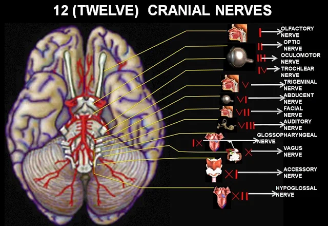
CRANIAL
NERVES:
1)
OLFACTORY NERVES (SENSORY):
These are
the nerve of sense of smell. Their sensory receptor and fibres originate
in the upper part of the mucous membrane of the nasal cavity.Pass upwards
through the cribriform plate of the ethmoid bone and then go to the olfactory
bulb.The nerve then proceed backwards as the olfactory tract,to the area
for the perception of smell in the temporal lobe of the cerebrum.
2) OPTIC
NERVES (SENSORY):
These are
the nerves of sense of sight.The fibres originate in the retinae of the
eyes and they combine to form the optic nerves.They are directed backward and
medially through the posterior part of the orbital cavity.They then pass
through the optic foramina of the sphenoid bone into the cranial cavity
and join at the optic chiasma.The nerves proceed back-wards as the optic
tracts to the lateral geniculate bodies of the thalamus.Impulses pass
from these to the centre for sight
in the
occipital lobes of the cerebrum and to the cerebellum.In the occipital lobe
sight is perceived,and in the cerebellumthe impulses from the eyes contribute
to the maintaenance of balance,posture and orientation of the head in space.
3)
OCULOMOTOR NERVES (MOTOR):
These
nerves arise from nuclei near the cerebral aqueduct.They supply:
·
Four of the six extrinsic muscles,which move
the eyeball, i.e. the superior,medial and inferior recti and inferior oblique muscle.
·
The intrinsic muscles: Ciliary muscle,which
alter the shape of lens,changing its refractive power. Circular muscle,of the iris,which contrict the pupil.
·
The levator palpebrae muscles, which raise
the upper eyelid.
4)
TROCHLEAR NERVES (MOTOR):
These nerves arise from nuclei near the
cerbral aqueduct. They supply the superior oblique muscles of the eyes.
5)
TRIGEMINAL NERVES (MIXED):
These
nerves contain motor and sensory fibres and are among the largest of the
cranial nerves.They are the chief sensory nerve for the faces and
head(Including the oral and nasal cavities and teeth) receiving impulses of
pain,temparature and touch.The motor fibres stimulate the muscles of
mastication. As name suggests,there are 3 main branches of the trigeminal
nerves. The dermetatomes innervated by the sensory fibres .
The opthalmic
nerve are sensory only and
supply the lacrimal glands,conjunctiva of the eyes,forehead,eyelids, anterior
aspect of the scalp and mucous membrane of the nose.The maxillary nerves
are sensory only and supply the cheeks ,upper gums,upper teeth and lower
eyelids.
The mandibular
nerves contain both sensory and motor fibres.These are the largest of
the 3 divisions and they supply the teeth and gums of the lower jaw,pinnae of
the ears lower lip and tongue.The motor fibres supply the muscles of
mastication.
6)
ABDUCENT NERVES (MOTOR):
These
nerves arise from nuclei lying under the floor of the 4th ventricle.They supply
the lateral rectus muscle of the eyeballs.
7) FACIAL
NERVES (MIXED):
The
nerves are composed of both motor and sensory nerve fibres,arising from nuclei
in the lower part of the pons.The motor fibres supply the muscles of the facial
expression.The sensory fibres convey impulses from the taste buds in the
anterior two third of the tongue of the taste perception area in the cerebral
cortex.
8)
VESTIBULOCOCHLEAR NERVES (AUDITORY)(SENSORY):
These
nerves are composed of two distinct sets of fibres,vestibular nerves and
cochlear nerves.
·
The Vestibular nerves arise
from the semicircular canals of the inner ear and convey impulses to the
cerebellum.
they are associated with the maintenance of posture and balance.
·
The Cochlear nerves originate
in the spiral organ in the inner ear and convey impulses to the hearing area in
the
cerebral cortex where sound is
perceived.
9)
GLOSSOPHARYNGEAL NERVES (MIXED):
The motor
fibres arise from nuclei in the medulla oblongata and stimulate the muscles of
the tongue and pharynx and the secretory cells of the parotid glands. The
sensory fibres convey impulses to the cerebral cortex from the posterior third
of the tongue. The tonsil and pharynx and from taste buds in the tongue and
pharynx.These nerves are essential for the swallowing and gag reflexes.
10) VAGUS
NERVES(MIXED):
These
nerves have a more extensive distribution than any other cranial nerves.They
pass down through the neck into the thorax and the abdomen.These nerves form an
important part of the parasympathetic nervous system.The motor fibres arise
from nucleiin the medulla and supply the smooth muscle and secretory glands of
the pharynx larynx ,trachea,heart, oesophagus,stomach ,intestine,pancreas ,
gall bladder, bile duct ,spleen ,kidney,ureter and blood vessels in the
thoracic and abdominal cavities.
11)
ACCESSORY NERVES(MOTOR):
These
nerves arise from nuclei in the medulla oblongata and in the spinal cord.The
fibres supply sternocleidomastoid and trapezius muscles. Branches join the
vagus nerves and supply the pharyngeal and laryngeal muscles.
12)
HYPOGLOSSAL NERVES (MOTOR):
These
nerves arise from nuclei in the medulla oblongata.They supply the muscles of
the tongue and muscles surrounding the hyoid bone and contribute to swallowing
and speech.
AUTONOMIC
NERVOUS SYSTEM:
The
autonomic or involuntary part of the nervous system controls the 'automatic'
functions of the body,i.e. initiated in the
brain
below the level of the cerebrum. Although stimulation does not occur
voluntarily, the individual may be conscious
of its
effects, e.g. an increase in their heart rate the effect of autonomic activity
are rapid and the effector organs rate:
·
Smooth muscle, e.g. changes in airway or
blood vessel diameter.
·
Cardiac muscle, e.g. changes in rate and
force of the heartbeat.
·
Glands, e.g. increasing or decreasing
gastrointestinal secretions.
The
efferent (motor) nerves of the autonomic nervous system arise from nerve cells
in the brain and emerge at various levels between the midbrain and the sacral
region of the spinal cord.Many of them travel within the same nerve sheath
as the
peripheral nerves of the central nervous system to reach the organs that they
innervate.
The autonomic nervous system is separated
into 2 division:-
·
Sympathetic
(Thoracolumbar outflow)
·
Parasympathetic (Craniosacral
outflow)
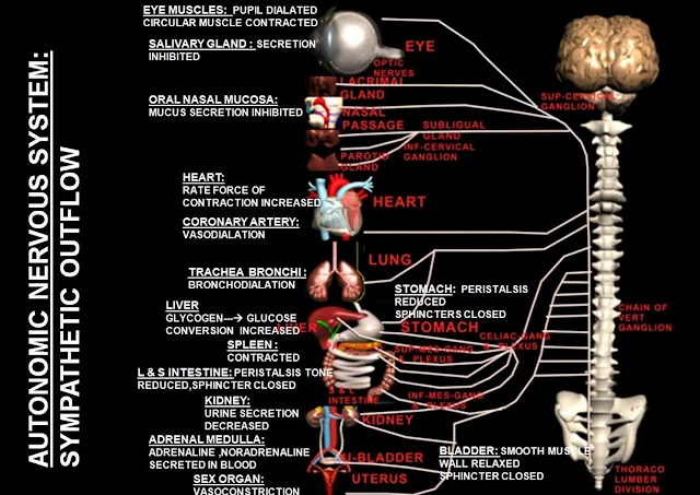
SYMPATHETIC
NERVOUS SYSTEM:
Neurones
impulses from their origin in the hypothalamus,reticular formation and medulla
oblongata to effector organs and tissues. The 1st neurone has its cell body in
the brain and its fibres extends into the spinal cord. The preganglionic and
post ganglionic neurones then conduct sympathetic impuses to effector organs.
The pre
Ganglionic neurone:
This has
its cell body in the lateral column of grey matter in the spinal cord between
the lavels of the 1st thoracic and 2nd or 3rd lumber vertebrae. The nerve
fibres of this cell leaves the cord by the anterior root and terminates at a
synapse in one of the ganglia either in the lateral chain of sympathetic
ganglia or passes through it to one of the pre vertebral ganglia. Acetylcholine
is the neuro transmitter at sympathetic ganglia
The Post
Ganglionic neurone:
This has
its cell body in a ganglion and terminates in the organs or tissue supply
noradrenaline is usually the neurotransmitters
at sympathetic effector organ.The major exception is that their is no
parasympathetic supply to the sweat glands,
the skin and blood vessel of skeletal muscles.This structure are supplyed by
only sympathetic post-ganglionic neuron
which usually has acetylcholine as their neurotransmitter they have therefore,
the effects of both sympatheticand
parasympathetic nerve supply.
There are 3 prevertebral ganglia situated
in the abdominal cavity close to the origins
of the
arteries of the same names:
·
COELIAC GANGLION:
·
SUPERIOR MESENTERIC GANGLION:
·
INFERIOR MESENTERIC GANGLION:
The
ganglia consists of nerve cell bodies rather diffusely distributed among a
network of nerve fibres that form plexuses
AUTONOMIC
NERVOUS SYSTEM:
The
autonomic or involuntary part of the nervous system controls the 'automatic'
functions of the body,i.e. initiated in the
brain
below the level of the cerebrum. Although stimulation does not occur
voluntarily, the individual may be conscious
of its
effects, e.g. an increase in their heart rate the effect of autonomic activity
are rapid and the effector organs rate:
·
Smooth muscle, e.g. changes in airway or
blood vessel diameter.
·
Cardiac muscle, e.g. changes in rate and
force of the heartbeat.
·
Glands, e.g. increasing or decreasing
gastrointestinal secretions.
The
efferent (motor) nerves of the autonomic nervous system arise from nerve cells
in the brain and emerge at various levels between the midbrain and the sacral
region of the spinal cord.Many of them travel within the same nerve sheath as the
peripheral nerves of the central nervous system to reach the organs that they
innervate.
The autonomic nervous system is separated
into 2 division:-
·
Sympathetic
(Thoracolumbar outflow)
PARASYMPATHETIC
NERVOUS SYSTEM:
Two
neurones (Preganglionic and postganglionic) are involved in the transmission of
impulses from their source to the
effector
organ.The neurotransmitter at both synapses is acetylcholine.
The
preganglionic neurone:
This is
ususally long in comparison to its counterpart in the sympathetic nervous
system and has its cell body either in the brain or in the spinal cord.Those
originating in the brain are the cranial nerves 3rd 4th 9th and 10th arising
from nuclei in the midbrain and brain stem, and their nerve fibres terminate
outside the brain.The cell bodies of the sacral ouflow are in the lateral
columns of grey matter at the distal end of the spinal cord.Their fibres laeve
the cord in sacral segments 2, 3 and 4 and ynapse with postganglionic neurones
in the walls of pelvic organs.
The
postganglionic neurone:
This is
usually very short and has its cell body either in a ganglion or, more often,
in the wall of the organ supplied.
 |
| Major Human Nerves |


 Online Movies
No comments:
Post a Comment