


 |
| Position and Different Organs of Digestive System |
DIGESTIVE SYSTEM:
The activities in the digestive system can be
grouped under 5 main heading.
1) INGESTION:
This is the taking of food into the
alimentary tract i.e. eating and drinking.
2) PROPULSION:
This mixes and moves the contents along the
alimentary tract.
3) DIGESTION:
This consist of :
a) Mechanical Breakdown of Food e.g : Mastication ( Chewing)
b) Chemical digestion of food into small
molecules by enzyme present in secretions produced by glands and accessory
organs of the digestive system.
4) ABSORPTION:
This is the process by which digested food
substances pass through the wall of some organs of the alimentary canal into
the blood and lymph capillaries for the circulation and use by body cell.
5) ELIMINATION:
Food subtances that have been eaten but
cannot be digested and absorbed are excreted from the alimentary canal as
faeces by the process of defaecation.
ORGANS OF THE DIGESTIVE
SYSTEM:
1) Alimentary tract:
Also known as gastrointestinal (GI) tract,
This is the long tube through which the food passes.It commences at the mouth
and terminates at anus,and the various parts are given separate names, although
structurally they are remarkably similar. The parts are:
a) MOUTH
b) PHARYNX
c) OESOPHAGUS
e) STOMACH
d) SMALL INTESTINE
d) SMALL INTESTINE
f) LARGE INTESTINE
g) RECTUM AND ANAL CANAL.
2) Accessory organs:
Various secretion are poured into the
alimentary tract, some by glands in the lining memebrane of the organs,e.g.
gastric juice secreted by glands in the lining of the stomach,and some by
glands situated outside the tract.The latter are the accessory organs of
digestion and their secretions pass through ducts to enter the tract.
They consist of:
a) 3 pairs of salivary glands
b) The liver and biliary tract.
The organs and glands are linked
physiologically as well as anatomically in that digestion and absorption occur
in stages,each stage being dependent upon previous stage or stages.
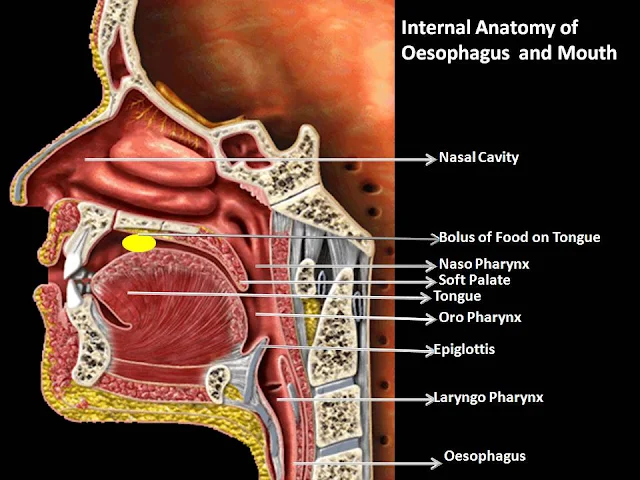 |
| Mouth and Oesophagus of Human with all Parts Name |
OESOPHAGUS
& MOUTH:
Structure:
there are 4 layers of
tissue , The oesophagus is almost entirely in the thorax the outer covering,the
adventitia,consist of elastic fibrous tissue that attaches the oesophagus to
the surrounding stuctures.The proximal third is lined by saritified squamous
epithelium and distal third by columnar epithelium.The middle third is lined by
mixture of two.
Function
of mouth pharynx and Oesophagus:
Formation of a Bolus:
When food is taken
into the mouth it is masticated,or chewed by the teeth and moved run the mouth
by the tongue and muscles of the cheeks.It is with saliva and formed into soft
mass or bolus ready for swallowing.The length of time that food remains in the
mouth depends. To a large extent, on the consistency of the food.Some foods
need to be chewed longer than others before the individual feels that the bolus
is ready for swallowing.
 |
| Mouth and Oesophagus of Human |
Swallowing(deglutition):
This occurs in 3
stages after mastication is complete and the bolus has been formed.It is
initiated voluntarily but completed by a reflex(involuntary) action.
1) The mouth is
closed and the voluntary muscles of the tongue and cheeks push the bolus
backwards into the pharynx.
2) The muscles of the
pharynx are stimulated by a reflex action initiated in the walls of the oropharynx and coordinated in the medulla and
lower pons in the brain stem.Involuntry contraction of these muscle propels the
bolus down into the oesophagus.All other routes that the bolus take are
closed.The soft palate rises up and closes of the nasopharynx;the tongue and
pharyngeal folds block the way back into the mouth; and larynx is lifted up and
forward so that its opening
is occluded by the
over hanging epiglottis preventing entry into the airway(trachea).
3) The presence of
the bolus in the pharynx stimulates a wave of peristalsis that propels the
bolus through the oesophagus to the stomach.
 |
| Human Stomach with its Part Names |
STOMACH:
The
stomach is a J-shaped dilated portion of the alimentary tract situated in the
epigastric,umbilical and left hypocondriac regions of the abdominal cavity.
Organs
associated with stomach:
Anteriorly-
Left lobe of liver and anterior abdominal wall
Posteriorly-
Abdominal aorta,pancreas,spleen,left kidney and adrenal gland.
Superiorly
- Diaphragm,oesophagus and left lobe of liver.
Inferiorly
- Transverse colon and small insestine.
Top to
left- Diaphragm and spleen.
To the
right- Liver and duodenum.
 |
| Human Stomach |
Structure
of stomach:
The
stomach is continuous with the oesophagus at the cardiac sphincter and with the
duodenum at the pyrolic sphincter it curves upward to complete the J
shape.Where the oesophagus join the stomach the anterior region angles acutely
upwards,curves downwards forming the great curvature and then slightly upwards
towards the pyrolic sphinter. The stomach is divided into 3 regions: The
fundus, The body and the antrum.At the distal end of the pyrolic antrum is the
pyrolic sphincter, guarding the opening between the stomach and the duodenum.When
the stomach is inactive the pyrolic sphincter is relaxed and open, and when the
stomach contains food the sphincter is closed.
Functions
of the stomach and gastric juice:
Stomach size varies
with the volume of food it contains,which may be 1.5 litres or more in an
adult. When a meal has been eaten the food accumulates in the stomach in
layers,the part of the meal remaining in the fundus for some time.Mixing with
the gastric juice takes place gradually and it
may be some time before the food is sufficiently acidified to stop the
action of salivary amylase. Gastric muscle contraction consists of a churning
movement that breaks down the bolus and mixes it with gastric juice and
peristaltic waves that propel the stomach contain towards the pyrolus.When the
stomach is active the pyrolic sphinter closes.Strong peristalic contraction of
the pyrolic antrum forces chyme,gastric contents after they sufficiently
liquefied,through the pyrolus into the duodenum in small spurts.Parasympathetic
stimulation increses the mortality of the stomach and secretion of the gastric
juice; sympathetic stimulation has the opposite effect.
Gastric
juice:
About 2 litres of
gastric juice are secreted daily by specialised secretory glands in the
mucosa consists of:
Water :
Further liquefies the food swallowed.
Mineral salts
Mucus secreted by
goblet cells in the glands and on the stomach surface.It prevent the mechanical
injury to the stomach by lubricating the contents.
Hydrochloric acid:
Acidifies the food and stops the action of salivary amylase,kills ingested
microbes,provides acid environment needed for effective digestion by
pepsin.
Intrinsic factor
Inactive enzyme
Precursors: pepsinogens secreted by chief cells in the glands.They begin the
digestion of protein,breaking them into smaller molecules.
SMALL AND
LARGE INTESTINE:
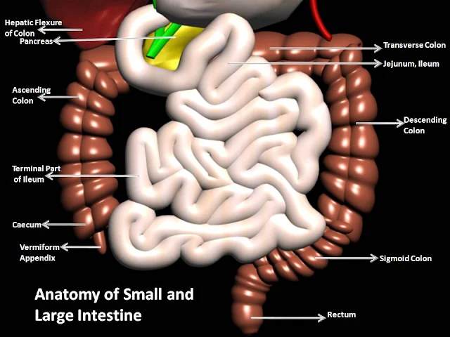 |
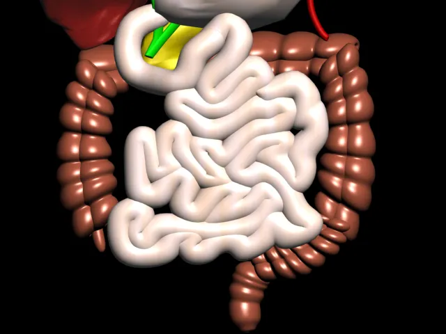
| Human Small and Large Intestine with its Parts Names |
The small intestine comprise 3 main sections continuos with each other.
1) Duodenum: it is
about 25 cm long and curves around the head of the pancreas secretion from the
gall bladder and pancreas are released into the duodenum through a common
structure the hepato-pancreatic ampulla and the opening into the duodenum is
guarded by the hepatopancreatic sphincter.
2) Jejunum: It is the
middle section of the small intestine and is about 2 metres long.
3) Ileum: The
terminal section,is about 3 metres long and ends at theileocaecal valve,which
cintrols the flow of material from the ileum to the caecum, the first part of
the large intestine and prevent regurgitation.
 |
| Human Small and Large Intestine |
Structure
of small intestine:
The walls of small
intestine are composed of the 4 layers of tissue
a) Peritoneum: A
double layer peritneum called the mesentry attaches the jejunum and ileum to
the posterior abdominal wall.
b) Mucosa: The suface
area of the small intestine mucosa is greatly incresed by permanent circular
folds,villi and microvilli.
Function
of the small intestine:
1) Upward movement of
its contents by peristalsis which is increased by parasympathetic stimulation.
2) Secretion of
intestinal juice,also increased by parasympathetic stimulation
3) Completion of
chemical digestion of carbohydrates protein and fats in the enterocytes of the
villi
4) Protection aganist
infection by microbes that have survived the antimicobial action of the
hydrochloric acid in the stomach,by the solitary lymph follicles and aggregated
lymph follicles.
5) Secretion of the
hormone cholecystokinin and secretin
6) Absorption of
nutrients.
 |
| Human Digestive System |
LARGE
INTESTINE:
This is about 1.5
metres long,beginning at the caecum in the right iliac fossa and terminating at
the rectum and anal canal deep in the pelvis. Its lumen is about 6.5 cm in
diameter, larger than that of the small intestine. The colon is divided into
the 5 parts
1)The caecum: The 1st
part of the colon. It is a dilated region which has a blind end inferiorly and
it is continuous with the ascending colon superiorly.
2)The ascending
colon: This passes upwards from the caecum to the level of the liver where it
curves acutely to the left at the hepatic flexure to become transverse colon.
3) Transverse colon:
This is the loop colon that extend
across the abdominal cavity in front of the duodenum and stomach to the
area of the spleen where it forms splenic flexure nad curves acutely downwards
to become the descending colon.
4) The descending
colon: This passes down the left side of the abdominal cavity then cuves
towards the midline.After it enters the true pelvis it is known as sigmoid
colon.
5) The sigmoid colon:
This part describes an s-shaped curve in the pelvis that continues downwards to
become the rectum.
6) Rectum: This
slightly dilated section of the colon about 13 cm long.Its leads from the
sigmoid colon about 13 cm long.It leads from the sigmoid colon and terminates
in the anal canal.
7) The anal canal:
This is a short passage about 3.8 cm long in the adult and leads from the
rectum to the exterior
 |
| Human Digestive System |
Functions
of the large intestine,rectum and anal canal:
a) Absorption: The
contents of the ileum which pass through the ileocaecal valve into the caecum
are fluid,even through some water has been absorbed in the small intestine.
b) Microbial
activity: The large intestine is heavily colonised by certain types of
bacteria,which synthesis vitamin K and Folic acid.They include E.coli,Enterobactor
aerogenes, Streptococcus faecalis, Clostridium perfringens.
c) Mass movement: The
large intestine does not exhibit peristaltic movement as in other parts of digestive tract.
d) Defaecation:
Usually the rectum is empty,but when the mass movement forces the contents of
the sigmoid colon into the rectum the nerve endings in its walls are stimulated
by strech.In the infants ,defaecation occurs by reflex(involuntary)
action.However,during the 2nd and 3rd year of the life the ability to override
the defaecation reflex is developed.
Constituents
of faeces:
Fibre(Indigestable
cellular plant and animal material) Dead and live microbes.
Epithelial cell shed
from the walls of tract Fatty acids Mucus secreted by epithelial linning of the
large intestine
 |
| Human Pancreas with its Part Names |
PANCREAS:
This pancreas is a
pale grey gland weighing about 60 grams.It is about 12 to 15 cm long and is
situated in the epigastric and left hypochondriac regions of the abdominal, It Consist fo a board head
and body and a narrow tail.
The head lies in the curve of a duodenum,The body behind the stomach and tail lies in front of the left kidney and just reaches the spleen.The abdominal aorta and the inferior vena cava lie behind the gland.It both an exocrine and endocrine gland.
LIVER:
The liver is the largest gland in the body, weighing between 1 and 2.3 kg.It is situated in the upper part of the abdominal cavity occupying the greater part of the right hypochondric region,part of the epigastric region and extending into the left hypochondriac region. Its upper and anterior surface is smooth and curved to fit the under surface of the diaphragm;its posterior surface is irregular in outline.
Organs associated with the liver:
Superiorly & anteriorly- Diaphragm and anterior abdominal wall.
Inferiorly- Stomach,bile duct,duodenum,hepatic fixture of colon right kidney and adrenal gland.
Posteriorly-Oesophagus,inferior vena cava,aorta,gall bladder,vertebral column and diaphragm.
Laterally- Lower ribs and diaphragm.
Liver enclosed in a thin inelastic capsule and incompletely covered by a layer of peritonium.Folds of peritoneum from supporting ligaments attaching the liver to the inferior surface of the diaphragm.It is held in position partly by these ligaments and partly by the pressure of the organs in the abdominal cavity.Liver has 4 lobes.The 2 most ovious are the large right lobe and the smaller,wedge-shaped,left lobe.The other 2,The caudate and quadrate lobes,are areas on the posterior surface.
Functions of the liver:
1) Carbohydrate Metabolism
2) Fat metabolism
3) Protein metabolism
4)Breakdown of erythrocytes and defence aganist microbes
5) Detoxification of drugs and noxious substances
6) Inactivation of hormones: This include insulin,glucagon,cortisol,aldosterone,thyroid and sex hormone.
7) Production of heat
8)Secretion of heat
9)Secretion of bile
10)Storage
 |
| Human Pancreas |
The head lies in the curve of a duodenum,The body behind the stomach and tail lies in front of the left kidney and just reaches the spleen.The abdominal aorta and the inferior vena cava lie behind the gland.It both an exocrine and endocrine gland.
 |
| Human Liver with its Parts Name |
LIVER:
The liver is the largest gland in the body, weighing between 1 and 2.3 kg.It is situated in the upper part of the abdominal cavity occupying the greater part of the right hypochondric region,part of the epigastric region and extending into the left hypochondriac region. Its upper and anterior surface is smooth and curved to fit the under surface of the diaphragm;its posterior surface is irregular in outline.
 |
| Anterior Part of The Human Liver |
Organs associated with the liver:
Superiorly & anteriorly- Diaphragm and anterior abdominal wall.
Inferiorly- Stomach,bile duct,duodenum,hepatic fixture of colon right kidney and adrenal gland.
Posteriorly-Oesophagus,inferior vena cava,aorta,gall bladder,vertebral column and diaphragm.
Laterally- Lower ribs and diaphragm.
Liver enclosed in a thin inelastic capsule and incompletely covered by a layer of peritonium.Folds of peritoneum from supporting ligaments attaching the liver to the inferior surface of the diaphragm.It is held in position partly by these ligaments and partly by the pressure of the organs in the abdominal cavity.Liver has 4 lobes.The 2 most ovious are the large right lobe and the smaller,wedge-shaped,left lobe.The other 2,The caudate and quadrate lobes,are areas on the posterior surface.
 |
| Posterior Part of The Human Liver |
Functions of the liver:
1) Carbohydrate Metabolism
2) Fat metabolism
3) Protein metabolism
4)Breakdown of erythrocytes and defence aganist microbes
5) Detoxification of drugs and noxious substances
6) Inactivation of hormones: This include insulin,glucagon,cortisol,aldosterone,thyroid and sex hormone.
7) Production of heat
8)Secretion of heat
9)Secretion of bile
10)Storage
 |
| Salivary Gland |
SALIVARY
GLANDS:
Its release their
secretions into ducts that lead to the mouth.There are 3 main pairs;the parotid
glands, the submandibular glands and the sublingual glands.There are also
numerous smaller salivary glands scattered around the mouth.
 |
| SALIVARY GLANDS |
Parotid
glands:
These are situated
one on each side of the face just below the external acoustic meatus. Each
gland has a parotid duct opening into the mouth at the level of the 2nd upper
molar tooth.
Submandibular
glands:
These lie on each
side of the face under the angle of the jaw.The 2 submandibular ducts open on
the floor of the mouth,one on each side of the frenulum of the tongue.
Sublingual
gland:
These glands lie
under the mucous membrane of the floor of the mouth in front of the
submandibular glands.They have numerous small ducts that open into the floor of
the mouth.
Composition
of saliva:
1.5 litres of saliva
is produced daily and it consist of:
Water
Mineral salts
Salivary amylase
mucus
Lysozyme
Immunoglobulins
Blood -clotting
factors.
Functions
of saliva:
1) Chemical digestion
of polysaccharides:
2) Lubrication of
foods
3) Cleaning and
lubricating
4) Non-specific
defence
5)Taste
 Nature Magazine
 National Geographic
 Cartoon Games
 History Channel
 Online Movies
 Scientific American
 Filmfare Mag
 Online Britannica
 Time Mag
 Unexplained Mysteries


No comments:
Post a Comment