USING WAVES
AND ELECTRONS:
 |
| Light Wave Page |
WAVES:
Light is
form of energy that enables us to see and makes plants grow. A light beam can
travel through empty space.So this energy does not use the air or other
material through which it passes in order to travel.The energy must must
therefore be carried by the beam itself.
We can
tell from the sharp edges of shadows
that rays of light travel through air along straight path.They cannot bend
round corners.
Animation showing different Wave Form of Light
People
therefore thought of light rays as straight lines. They help to explain
reflection and refraction. Then in 1680,Huygens suggested that light rays were
in fact waves. Over a century later his theory was shown to be true.
WAVE LENGTHS:
 |
| Different Wave Forms of Light |
Light
travels in one direction but the ray itself is moving up and down in continuous
crests and troughs.This wave has a similar shape to ripples on a pond.As it
moves through space,there is always the same distance between two neighbouring
crests or troughs. The distance is called the wavelength.It is an extremely
tiny distance measured in minute fractions of a meter.The height of a crest or
the depth of a trough is called the amplitude.The greater the amplitude of the
wave, the greater its energy.As the energy decreases, the amplitude grows less
and less.
 |
| Graphical Representation Wavelength and Direction of Light Wave |
After the
wave has gone through one crest and one trough it has travelled one
wavelength.The wave has completed one cycle of its motion and is read to
repeat itself.The number of cycles in one second is called the frequency of the
wave.
Waves
move at tremendous speed.The speed is always the same in one particular medium
such as air,but decreases when waves enter denser material,such as glass or
water.This change in speed causes refraction of the light beam producing an
increase in wavelength.The speed of a light wave in any medium equals its
wavelength multiplied by its frequency . The greatest speed of light is in a
vacuum,such as outer space .The speed in air is very close to this value.The
maximum speed is equal to 300,000 Km per second .No object moving in a vacuum
can travel faster than this speed.
Each
light wave has its own wavelength and each of these wavelengths corresponds to
a slightly different color.Red light has almost twice the wavelength of violet
light .Yellow.green and blue light have wavelengths between these values.
ELECTROMAGNETIC
RADIATION:
 |
| Visible Light Spectrum |
Light is
not the only form of energy transmitted in waves , Radio waves, infrared and
ultraviolet radiation,X-ray and gamma rays also travel as wave motions.All
these waves move at the speed of light .However,the wave lengths (and hence
frequencies) are very different. It is the different wavelengths that give each
type of radiation its special properties.They are
all examples of electromagnetic radiation, and they all travel as
electromagnetic waves.
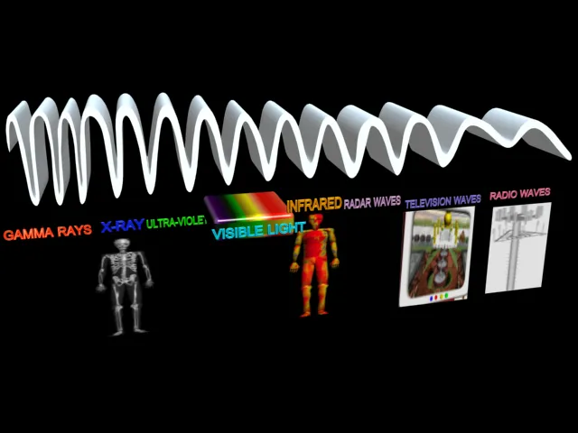
The chart
showing the different electromagnetic radiations in order of increasing
wavelength (or decreasing frequency) is called the electromagnetic spectrum.
 |
| Graphical Representation of Different wave length of Visible colored light |
Wave
lengths of Red light is almost twice that of blue light .The
frequency is that the number of complete wave cycle in one second. As the
wavelength increases the frequency decreases .Red light has almost twice the
wavelength of blue light while its frequency is almost half that of blue light.
 |
| Graphical relation of light wave energy and amplitude |
The
amplitude or maximum height of a crest
or trough,remains the same if the wave`s energy stays the same .If the
wave loses the
energy , the amplitude decreases. The square of the amplitude (amplitude times
amplitude)gives a measure of the energy.
|
| Visible Light Spectrum |
The electromagnetic
spectrum. Radio waves have much longer wavelengths than light waves which in
turn have greater wavelengths than X-rays and gamma rays .We can only see a
very narrow part of the spectrum.
 |
| X-Ray Page |
X-RAYS:
 |
Wilhelm Conrad Röntgen : was a German physicist, who, on 8 November 1895, produced and detected electromagnetic radiation in a wavelength range today known as X-rays
|
Animation Showing X-Ray Machine and its Inner Technology
Like
light, X-rays also move as waves, but
the wavelength is very much smaller. Unlike light waves, they are invisible.The
energy of X-rays is very high. They can travel a great distance inside an
object and sometimes they are able to pass straight through it.
 |
| 3D Picture of X-Ray Tube and it components |
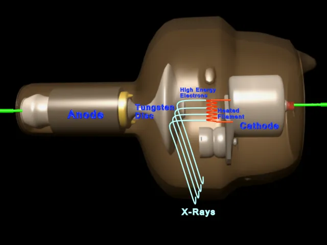
X-rays
are produced in an X-ray tube when a narrow stream of electrons emitted from a
heated cathode is strongly attracted towards the anode.The anode is at a very
high voltage.This means that the electrons move very fast towards the anode and
therefore have a large amount of energy.The anode contains a small disc of
heavy metal such as Tungsten.When the electrons strike the tungsten atoms, They
give up their energy to electrons in the atoms.To get rid of this excess energy
, the atoms emit X-rays.
 |
| Full Picture of X-Ray Machine |
 |
| Close View of X-ray machine and Tube |
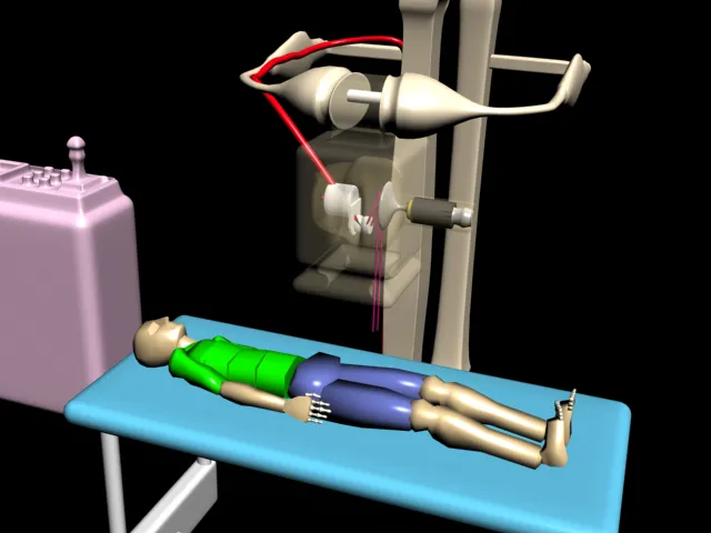
X-rays
have various uses in medicine X-rays, like light, acn produce an image on
photographic film .If a person stand between a low energy source of X-rays and
a film , a photo of the bone structure is obtained .Broken or badly formed
bones can be seen.
 |
| X-Ray Picture of Full Human Body |
CT-SCANNER:
 |
| Picture of CT-Scanner |
A more
advanced type of X-ray machine is the computerized tomographic (CT) scanner. A
CT scanner uses a computer to be produce highly detailed pictures of the inside
of a patient`s body. The computer in a CT machine is fed data on how the
different tissues in the body absorb X-rays.As a patient is scanned , the
computer compares this data with the amount of radiation actually absorbed by
the patient`s body.The computer is able to build up a very detailed picture of
the tissues in the body.
To Produce a CT scan ,X-rays are only passed through a thin slice of the body at
one time .This also helps produce a clearer picture .The X-ray tube travels
around the patient`s body , making 1.5 million exposures as it travels.
Sensitive detectors also travel around
the body opposite the X-ray tube.The detectors are linked to the computer which
builds up the detailed picture on a television -like screen.Today`s scanners
can examine slices of the body from 2 mm to 13 mm thick,as easily as if they
were slices of bread.
NMR
SCANNER:
Another
type of body scanner is called an NMR scanner.This uses a process called
nuclear magnetic resonance (NMR) to
produce detailed pictures of the inside of a patient`s body.The patient lies in a strong magnetic field produced by
an electric current flowing in a superconducting coil.Radio signals are beamed
into the area of the body being investigated .The
nuclei or central parts, of the atoms of the body produced magnetic signals
which are picked up by detectors. A computer is used to form a picture of the
inside of the body from the magnetic signals.
 |
| Picture of MRI Scanner |
X-rays
are also used in scientific research to find out how atoms and molecules are
arranged in crystals and how they are grouped together in some of then giant
chemical compounds found in the body,such as DNA.This type of research is
called x-ray crystallography.
 |
| 3D Picture of Diode as Rectifier |
 |
| Different Parts of Diode |
Animation on Diode
The diode
. An alternating voltage makes the anode positive and then
negative in one cycle as current will only flow when the anode is positive ,
the negative half of the cycle is lost .Alternating current is thus changed to
direct current .The diode therefore acts as a rectifier.
 |
| 3D Picture of X-Ray Tube |
 |
| Elementary 2D picture of X-Ray Tube |
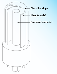
3-D
diagram of an x-ray tube. The cathode is specially
shaped so that a narrow beam of electrons is directed onto the tungsten
disc in the anode .This produces a fairly narrow beam of X-rays which leaves
the tube through a thin metal plate .The energy of the X-rays depends on the
difference in voltage between anode and cathode.
 |
Wilhelm Conrad
Röntgen : was a German physicist, who, on 8 November 1895, produced and
detected electromagnetic radiation in a wavelength range today known as
X-rays.
|
An X-ray
photograph of a hand with a ring .Dentists take X-ray photos to
check that your teeth are growing correctly and to see if you need any
fillings. Only bones and show up on the negative of the film. The film is
blackened by X-rays which are able to pass straight through the skin and air . Bones and teeth
absorb most of the X-rays and therefore appear whitish on the negative and dark
on the print.
The
chemical structure of cytochrome C. This is the giant molecule
present in the cells of the body .Its complicated structure was unravelled by
the use of X-rays. Each colored ball represent a different group of atoms
.Knowing its structure helps scientist to learn about how it works.
 |
| Computer Generated 3D Model of Cytochrome C Through X-Ray Crystallography Technique |
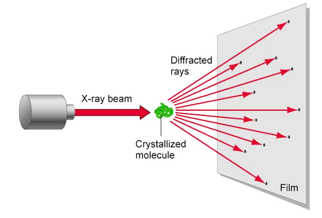 |
| X-Ray Crystallography Technique require to create a 3d Model in computer of any complex protein structure |


 Nature Magazine
 National Geographic
 Cartoon Games
 History Channel
 Online Movies
 Scientific American
 Filmfare Mag
 Online Britannica
 Time Mag
 Unexplained Mysteries


No comments:
Post a Comment