


All Eukaryotic cells usually are Two Types 1) Animal Cells 2) Plant Cells.
Animal cells :
are Eukaryotic cells, or cells with a membrane-bound nucleus. Unlike Prokaryotic cells, DNA in animal cells is housed within the nucleus.
In addition to having a nucleus, animal cells also contain other membrane-bound organelles, or tiny cellular structures, that carry out specific functions necessary for normal cellular operation. Organelles have a wide range of responsibilities that include everything from producing hormones and enzymes to providing energy for animal cells.
Animal Cells: Structures and Organelles
The following are examples of structures and organelles that can be found in typical animal cells:
Centriole - organize the assembly of microtubule during cell division.
Cytoplasm - gel-like substance within the cell.
Endoplasmic Reticulum - extensive network of membranes composed of both regions with ribosomes (rough ER) and regions without ribosomes (smooth ER).
Golgi Complex - responsible for manufacturing, storing and shipping certain cellular products.
Lysosomes - sacs of enzymes that digest cellular macromolecules such as nucleic acids.
Microtubules - hollow rods that function primarily to help support and shape the cell.
Mitochondria - power producers and the sites of cellular respiration.
Nucleus - membrane bound structure that contains the cell's hereditary information.
Nucleolus - structure within the nucleus that helps in the synthesis of ribosomes.
Nucleopore - tiny hole within the nuclear membrane that allows nucleic acids and proteins to move into and out of the nucleus.
Ribosomes - consisting of RNA and proteins, ribosomes are responsible for protein assembly.
1) Cytoskeleton:
This consists of tiny strands of protein.
Microfilaments:
These are the smallest fibres.They are provide structural support, maintain the characteristic shape of the cell and permit contraction e.g. in muscle cells.
Microtubules:
These are larger contractile protein fibres that are involved in movement of
Organelles within the cell
Chromosomes during cell division
Cell extensions
Centrosome:
This directs organisation of microtubules within the cell.It consists of a pair of centrioles (Small clusters of microtubules)
and plays an important role during cell division.
Cell Extensions:
These project from the plasma membrane and their main components are microtubules, which allow movement.They
include:
Cilia Small hair like projections lying along the free border of some cells.They beat in unison moving substances
along the surface e.g. mucus upwards in the respiratory tract.
Flagella Single, long whip-like projections that move cells, e.g. tails of spematozoa.
2) Mitochondria:
are the cell's power producers. They convert energy into forms that are usable by the cell. Located in the cytoplasm, they are the sites of cellular respiration which ultimately generates fuel for the cell's activities. Mitochondria are also involved in other cell processes such as cell division and growth, as well as cell death.
Mitochondria are bounded by a double membrane. Each of these membranes is a phospholipid bilayer with embedded proteins. The outermost membrane is smooth while the inner membrane has many folds. These folds are called cristae. The folds enhance the "productivity" of cellular respiration by increasing the available surface area.
The double membranes divide the mitochondrion into two distinct parts: the inter membrane space and the mitochondrial matrix. The intermembrane space is the narrow part between the two membranes while the mitochondrial matrix is the part enclosed by the innermost membrane. Several of the steps in cellular respiration occur in the matrix due to its high concentration of enzymes.
Mitochondria are semi-autonomous in that they are only partially dependent on the cell to replicate and grow. They have their own DNA, ribosomes and can make their own proteins. Similar to bacteria, mitochondria have circular DNA and replicate by a reproductive process called fission.
3) Golgi Apparatus:
This consist of stacks of closely folded flattened membranous sacs.It is present in all cells but is larger in those that systhesis and export proteins.The proteins move from the endoplasmic reticulum to the Golgi apparatus where they are packed into membrane bound vesicles called secretory granules . The vesicles are stored and, when needed, move to plasma membrane, through which the proteins are exported.
4) Ribosomes:
These are tiny granules composed of RNA and protein.They synthesis proteins from amino acids, using RNA as the template.When present in free units or on small clusters in the cytoplasm,the ribosomes make proteins for use within the cell.These include the enzymes required for metabolism.Metabolic pathways consist of series of steps, each driven by a specific enzyme.Ribosomes are also found on the outer surface of the nuclear envelope and rough endoplasmic
reticulum where they manufacture proteins for export the cell.
5) Lysosomes:
Lysosomes are one type of the secretory vesicle formed by the Golgi apparatus.They contain a variety of enzymes involved in breaking down fragments of organelles and large molecules (RNA, DNA, Carbohydrates,Proteins) inside the cell into smaller particles that are either recycled, or extruded from the cell as waste material. Lysosomes in white blood cells contain enzymes that digest foreign material such as microbes.
6) Endoplasmic Reticulum(ER):
This is the series of interconnecting membranous canals in the cytoplasm. There are two types: smooth and rough.Smooth ER synthesise lipids and steroid hormones,and is also associated with the detoxification of some drugs.Rough ER is studded with ribosomes.These are the site of synthesis of proteins that are exported from cells.i.e.enzymes and hormones that are extruded from the parent cell to be used by cells elsewhere.
7) Vacuole
is a membrane-bound sac that plays roles in intracellular digestion and the release of cellular waste products. In animal cells, vacuoles are generally small.
8) Cell Membrane structure and function:
1) Cell membranes are composed of lipids(Phospholipids especially) and protein.
2) Membrane lipids are hydrophilic heads and hydrophobic tails and ehen surrounded by water they assemble spontaneously into a bilayer. All heads are at the 2 outer faces of a lipid bi-layer and all tails are sandwiched between them.
3) The lipid bilayer is the basic structure of all cell-membranes and serves as hydrophobic barrier between two solutions.
A plasma membrane separates the cytoplasm and extracellular fluid, and organelle membranes separate different solutions in the space of cytoplasm.
4) Membrane fluidity arises through rapid movements and packing variations among individual lipid molecules.
5) Membrane functions are carried out largely by proteins embedded in the bilayer or positioned at one or the other membrane surface.
6) Channel protein allow passage of water soluble subtances across the lipid bi-layer.
7) Tranport protein also allow passage of subtances across the bilayer.Unlike the channel protein they require the energy to pump specific substances into specific direction.
8) Electron transfer proteins accept electrons from one molecule and transfer them to another .In some cases, they also transfer H+ ions.
9) Recognition proteins function in tissue formation and later in cell to cell interactions. In multicelled animals the polysaccharide chains of recognition proteins form a sugar coat at the outer surface of plasma membrane.
10) Receptor proteins are like switches that are turned on or turned off when particular substances bind to them.They are specialized receivers of outside information that can trigger alterations in metabolism or cell behavior.
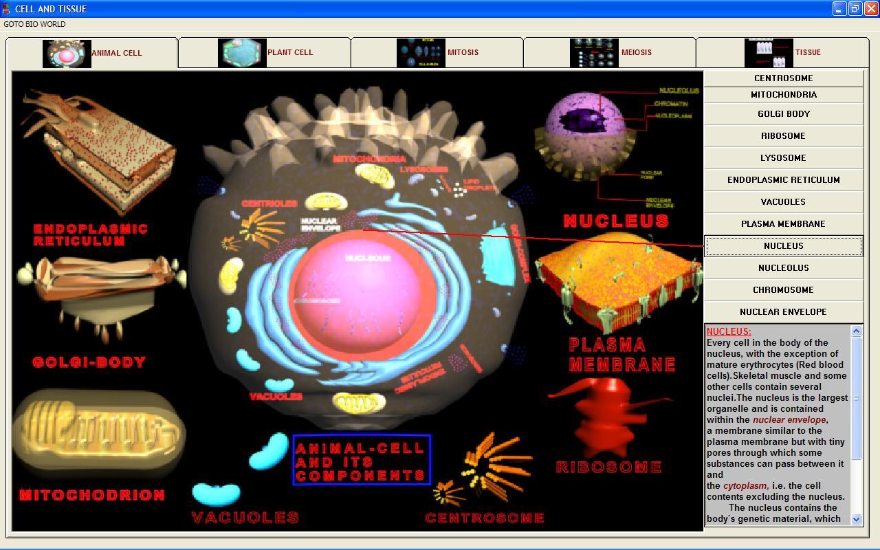
9) Nucleus:
Every cell have nucleus in the cell-body, with the exception of mature Erythrocytes (Red Blood Cells). Skeletal muscle and some other cells contain several nuclei. The nucleus is the largest organelle and is contained within the Nuclear envelope, a membrane similar to the plasma membrane but with tiny pores through which some substances can pass between it and the cytoplasm, i.e. the cell contents excluding the nucleus. The nucleus contains the body`s genetic material, which directs all thye metabolic activities of the cell. This consists of 46 chromosomes, which are made from deoxyribonucleic acid (DNA). Except during cell division, the chromosomes resemble a fine network of threads called chromatin.
10) Nucleolus:
As cells grow and develop, dense masses of irregular size and shape form in the nucleoplasm.They consist largely of RNA and proteins.Two or three masses form in most cells,but there can be a thousand or more in cells that develop into amphibian eggs.Each mass is a nucleolus, and it retains its distinct appearance only in nondividing cells.
This is the site where subunits of ribosomes are assembled.It is located on chromosomal regions that contain the instructions for synthesizing certain types of RNA molecules. Here, RNA is assembled, processed, and packaged with proteins into ribosomal subunit, which are then shipped to destinations in the cytoplasm. A complete, two-part ribosome is never found in the nucleus.
11) Nuclear Envelope:
This two-membrane envelope is the outermost component of the nucleus. Ribosome are attached to its outer surface, which faces the cytoplasm, and pores extent across the envelope at regular intervals.The structure of the nuclear envelope suggest that it is the boundary for controlled exchanges between the nucleus and the cytoplasm, with its pores being passageways.
Plant cells:
Plant Cells are eukaryotic cells that differ in several key respects from the cells of other eukaryotic organisms. Their distinctive features from Animal Cell include:
1) CENTRAL VACUOLES:
Mature and still living plant cells often have a large, fluid-filled central-vacuole. This organelle forms when small vesicles fuse during cell growth and development.It usually occupies as much as fifty to ninety percent of the cell interior, and this leaves only a narrow zone of cytoplasm between the vacuole`s membrane and the cell wall.
A central vacuole can serve as a storage area for metabolic products (such as amino acids and sugars), ions and toxic wastes.However, its main function may be to increase cell size and surface area.Water pressure builds up inside the vacuole to press against the cell wall. The cell enlarges permanently under this force.The extent of its enlargement depends partly on the volume possible within the confines of the wall, which is pliable at first but then become rigid at maturity.The increased surface area enhances the rate of absorption of the mineral salts necessary for plant nutrition,
A large central vacuole, a water-filled volume enclosed by a membrane known as the tonoplast maintains the cell's turgor, controls movement of molecules between the cytosol and sap, stores useful material and digests waste proteins and organelles.
2) CELL WALL:
For many cells, surface deposits outside the plasma-membrane form coats, capsules, sheaths and walls although some secrete products to the surface layer of tissues in which they occur.
Most cell walls have carbohydrate frameworks. They generally provide support and resist mechanical pressure, as when they keep plant cells from stretching too much while they expand with incoming water during growth.Even the most solid looking walls have microscopic spaces that make them porous, so water and solutes can move to and from the plasma-membrane.
In new plant cells, cellulose strands are bundled together and added to a developing "Primary" cell wall.After the main growth phase, many types of plant cells also deposit materials to form an inner, rigid "Secondary" cell wall, which often consist of several layers. Cutin, suberin and waxes commonly are embedded in cell walls at the plant`s surface; they play a protective role and help reduce water loss.
A cell wall composed of cellulose and hemicellulose, pectin and in many cases lignin, is secreted by the protoplast on the outside of the cell membrane. This contrasts with the cell walls of fungi (which are made of chitin), and of bacteria, which are made of peptidoglycan.
3) CHLOROPLAST STRUCTURE AND FUNCTION:
The light dependent reactions of photosynthesis take place at specialised cell membranes,such as Thylakoid membrane inside chloroplasts.That membrane is folded into the system pf stacked disks, called grana, and flatten channels.The interior spaces of the disks achannels are open to one another, so the membrane system actually forms a single compartment.The compartment is a reservoir for hydrogen ions, and it is tapped for ATP formation.The synthesis of phosphorylated sugars and starch occurs in the stroma, a semifluid matrix that surrounds the thylakoid compartment.
The internal organisation of a chloroplast might seem to be less than memorable, until you remember that we are taking about a very small space. If you could lined up 2000 chloroplasts, one after another, the lineup would be no wider than your thumbnail.Imagine all the chloroplasts in one lettuce leaf, each a tiny factory of producing sugars and starch and you begin to get a sense of the magnitude of metabolic events required to feed you and all other organism on earth.
Plastids, the most notable being the chloroplasts, which contain chlorophyll a green coloured pigment which is used for absorbing sunlight and is used by a plant to make its own food in the process is known as photosynthesis. Other types of plastid are the amyloplasts, specialized for starch storage, elaioplasts specialized for fat storage, and chromoplasts specialized for synthesis and storage of pigments. As in mitochondria, which have a genome encoding 37 genes, plastids have their own genomes of about 100–120 unique genes and, it is presumed, arose as prokaryotic endosymbionts living in the cells of an early eukaryotic ancestor of the land plants and algae.
4) Specialised cell–cell communication
pathways known as plasmodesmata, pores in the primary cell wall through which the plasmalemma and endoplasmic reticulum of adjacent cells are continuous.
Cytoplasm :
is basically the substance that fills the cell. It is a jelly-like material that is eighty percent water and usually clear in color. It is more like a viscous (thick) gel than a watery substance, but it liquefies when shaken or stirred. Cytoplasm, which can also be referred to as cytosol, means cell substance. This name is very fitting because cytoplasm is the substance of life that serves as a molecular soup in which all of the cell's organelles are suspended and held together by a fatty membrane. The cytoplasm is found inside the cell membrane, surrounding the nuclear envelope and the cytoplasmic organelles.
Animal cells :
 |
| 3D Picture of Animal Cell |
are Eukaryotic cells, or cells with a membrane-bound nucleus. Unlike Prokaryotic cells, DNA in animal cells is housed within the nucleus.
 |
| 3D Microscopic Picture of Animal Cell |
In addition to having a nucleus, animal cells also contain other membrane-bound organelles, or tiny cellular structures, that carry out specific functions necessary for normal cellular operation. Organelles have a wide range of responsibilities that include everything from producing hormones and enzymes to providing energy for animal cells.
 |
| Animal Cell Page in my Biology-World Software |
Animal Cells: Structures and Organelles
 |
| 3D Picture of Animal Cell and its Major Components |
The following are examples of structures and organelles that can be found in typical animal cells:
 |
| 3D Picture of Animal Cell and its Major Components |
Centriole - organize the assembly of microtubule during cell division.
Cytoplasm - gel-like substance within the cell.
Endoplasmic Reticulum - extensive network of membranes composed of both regions with ribosomes (rough ER) and regions without ribosomes (smooth ER).
Golgi Complex - responsible for manufacturing, storing and shipping certain cellular products.
Lysosomes - sacs of enzymes that digest cellular macromolecules such as nucleic acids.
Microtubules - hollow rods that function primarily to help support and shape the cell.
Mitochondria - power producers and the sites of cellular respiration.
Nucleus - membrane bound structure that contains the cell's hereditary information.
Nucleolus - structure within the nucleus that helps in the synthesis of ribosomes.
Nucleopore - tiny hole within the nuclear membrane that allows nucleic acids and proteins to move into and out of the nucleus.
Ribosomes - consisting of RNA and proteins, ribosomes are responsible for protein assembly.
1) Cytoskeleton:
This consists of tiny strands of protein.
Microfilaments:
These are the smallest fibres.They are provide structural support, maintain the characteristic shape of the cell and permit contraction e.g. in muscle cells.
Microtubules:
These are larger contractile protein fibres that are involved in movement of
Organelles within the cell
Chromosomes during cell division
Cell extensions
 |
| Centrosome |
Centrosome:
This directs organisation of microtubules within the cell.It consists of a pair of centrioles (Small clusters of microtubules)
and plays an important role during cell division.
Cell Extensions:
These project from the plasma membrane and their main components are microtubules, which allow movement.They
include:
Cilia Small hair like projections lying along the free border of some cells.They beat in unison moving substances
along the surface e.g. mucus upwards in the respiratory tract.
Flagella Single, long whip-like projections that move cells, e.g. tails of spematozoa.
2) Mitochondria:
 |
| Mitochondria |
are the cell's power producers. They convert energy into forms that are usable by the cell. Located in the cytoplasm, they are the sites of cellular respiration which ultimately generates fuel for the cell's activities. Mitochondria are also involved in other cell processes such as cell division and growth, as well as cell death.
Mitochondria are bounded by a double membrane. Each of these membranes is a phospholipid bilayer with embedded proteins. The outermost membrane is smooth while the inner membrane has many folds. These folds are called cristae. The folds enhance the "productivity" of cellular respiration by increasing the available surface area.
 |
| Mitochondria inner Structure |
The double membranes divide the mitochondrion into two distinct parts: the inter membrane space and the mitochondrial matrix. The intermembrane space is the narrow part between the two membranes while the mitochondrial matrix is the part enclosed by the innermost membrane. Several of the steps in cellular respiration occur in the matrix due to its high concentration of enzymes.
Mitochondria are semi-autonomous in that they are only partially dependent on the cell to replicate and grow. They have their own DNA, ribosomes and can make their own proteins. Similar to bacteria, mitochondria have circular DNA and replicate by a reproductive process called fission.
 |
| 3D Picture of Golgi apparatus |
3) Golgi Apparatus:
This consist of stacks of closely folded flattened membranous sacs.It is present in all cells but is larger in those that systhesis and export proteins.The proteins move from the endoplasmic reticulum to the Golgi apparatus where they are packed into membrane bound vesicles called secretory granules . The vesicles are stored and, when needed, move to plasma membrane, through which the proteins are exported.
 |
| Microscopic 3D picture of Ribosome |
4) Ribosomes:
These are tiny granules composed of RNA and protein.They synthesis proteins from amino acids, using RNA as the template.When present in free units or on small clusters in the cytoplasm,the ribosomes make proteins for use within the cell.These include the enzymes required for metabolism.Metabolic pathways consist of series of steps, each driven by a specific enzyme.Ribosomes are also found on the outer surface of the nuclear envelope and rough endoplasmic
reticulum where they manufacture proteins for export the cell.
 |
| Lysosome : 3D structure |
5) Lysosomes:
Lysosomes are one type of the secretory vesicle formed by the Golgi apparatus.They contain a variety of enzymes involved in breaking down fragments of organelles and large molecules (RNA, DNA, Carbohydrates,Proteins) inside the cell into smaller particles that are either recycled, or extruded from the cell as waste material. Lysosomes in white blood cells contain enzymes that digest foreign material such as microbes.
 |
| Endoplasmic Reticulum(ER) |
6) Endoplasmic Reticulum(ER):
This is the series of interconnecting membranous canals in the cytoplasm. There are two types: smooth and rough.Smooth ER synthesise lipids and steroid hormones,and is also associated with the detoxification of some drugs.Rough ER is studded with ribosomes.These are the site of synthesis of proteins that are exported from cells.i.e.enzymes and hormones that are extruded from the parent cell to be used by cells elsewhere.
 |
| Vacuole: 3D structure |
7) Vacuole
is a membrane-bound sac that plays roles in intracellular digestion and the release of cellular waste products. In animal cells, vacuoles are generally small.
 |
| Cell Membrane structure: 3D Structure |
8) Cell Membrane structure and function:
 |
| Cell Membrane structure picture |
1) Cell membranes are composed of lipids(Phospholipids especially) and protein.
2) Membrane lipids are hydrophilic heads and hydrophobic tails and ehen surrounded by water they assemble spontaneously into a bilayer. All heads are at the 2 outer faces of a lipid bi-layer and all tails are sandwiched between them.
3) The lipid bilayer is the basic structure of all cell-membranes and serves as hydrophobic barrier between two solutions.
A plasma membrane separates the cytoplasm and extracellular fluid, and organelle membranes separate different solutions in the space of cytoplasm.
4) Membrane fluidity arises through rapid movements and packing variations among individual lipid molecules.
5) Membrane functions are carried out largely by proteins embedded in the bilayer or positioned at one or the other membrane surface.
6) Channel protein allow passage of water soluble subtances across the lipid bi-layer.
7) Tranport protein also allow passage of subtances across the bilayer.Unlike the channel protein they require the energy to pump specific substances into specific direction.
8) Electron transfer proteins accept electrons from one molecule and transfer them to another .In some cases, they also transfer H+ ions.
9) Recognition proteins function in tissue formation and later in cell to cell interactions. In multicelled animals the polysaccharide chains of recognition proteins form a sugar coat at the outer surface of plasma membrane.
10) Receptor proteins are like switches that are turned on or turned off when particular substances bind to them.They are specialized receivers of outside information that can trigger alterations in metabolism or cell behavior.
 |
| 3D structure of Nucleus |
9) Nucleus:
Every cell have nucleus in the cell-body, with the exception of mature Erythrocytes (Red Blood Cells). Skeletal muscle and some other cells contain several nuclei. The nucleus is the largest organelle and is contained within the Nuclear envelope, a membrane similar to the plasma membrane but with tiny pores through which some substances can pass between it and the cytoplasm, i.e. the cell contents excluding the nucleus. The nucleus contains the body`s genetic material, which directs all thye metabolic activities of the cell. This consists of 46 chromosomes, which are made from deoxyribonucleic acid (DNA). Except during cell division, the chromosomes resemble a fine network of threads called chromatin.
10) Nucleolus:
As cells grow and develop, dense masses of irregular size and shape form in the nucleoplasm.They consist largely of RNA and proteins.Two or three masses form in most cells,but there can be a thousand or more in cells that develop into amphibian eggs.Each mass is a nucleolus, and it retains its distinct appearance only in nondividing cells.
This is the site where subunits of ribosomes are assembled.It is located on chromosomal regions that contain the instructions for synthesizing certain types of RNA molecules. Here, RNA is assembled, processed, and packaged with proteins into ribosomal subunit, which are then shipped to destinations in the cytoplasm. A complete, two-part ribosome is never found in the nucleus.
 |
| 3D Picture of Nucleus |
11) Nuclear Envelope:
This two-membrane envelope is the outermost component of the nucleus. Ribosome are attached to its outer surface, which faces the cytoplasm, and pores extent across the envelope at regular intervals.The structure of the nuclear envelope suggest that it is the boundary for controlled exchanges between the nucleus and the cytoplasm, with its pores being passageways.
 |
| Microscopic Picture slide of Blood Cell , Picture Taken by Me (Manash Kundu) |
 |
| Microscopic Picture slide of Kidney of mammal Picture Taken by Me (Manash Kundu) |
Plant cells:
 |
| 3D Picture of Plant Cell |
Plant Cells are eukaryotic cells that differ in several key respects from the cells of other eukaryotic organisms. Their distinctive features from Animal Cell include:
 |
| 3D Picture of Plant Cell and its Major Components |
 |
| Plant Cell Page in my Biology-World Software |
1) CENTRAL VACUOLES:
 |
| Anatomy of Plant Cell |
Mature and still living plant cells often have a large, fluid-filled central-vacuole. This organelle forms when small vesicles fuse during cell growth and development.It usually occupies as much as fifty to ninety percent of the cell interior, and this leaves only a narrow zone of cytoplasm between the vacuole`s membrane and the cell wall.
A central vacuole can serve as a storage area for metabolic products (such as amino acids and sugars), ions and toxic wastes.However, its main function may be to increase cell size and surface area.Water pressure builds up inside the vacuole to press against the cell wall. The cell enlarges permanently under this force.The extent of its enlargement depends partly on the volume possible within the confines of the wall, which is pliable at first but then become rigid at maturity.The increased surface area enhances the rate of absorption of the mineral salts necessary for plant nutrition,
A large central vacuole, a water-filled volume enclosed by a membrane known as the tonoplast maintains the cell's turgor, controls movement of molecules between the cytosol and sap, stores useful material and digests waste proteins and organelles.
2) CELL WALL:
 |
| Plant Cell With Cell Wall |
For many cells, surface deposits outside the plasma-membrane form coats, capsules, sheaths and walls although some secrete products to the surface layer of tissues in which they occur.
Most cell walls have carbohydrate frameworks. They generally provide support and resist mechanical pressure, as when they keep plant cells from stretching too much while they expand with incoming water during growth.Even the most solid looking walls have microscopic spaces that make them porous, so water and solutes can move to and from the plasma-membrane.
In new plant cells, cellulose strands are bundled together and added to a developing "Primary" cell wall.After the main growth phase, many types of plant cells also deposit materials to form an inner, rigid "Secondary" cell wall, which often consist of several layers. Cutin, suberin and waxes commonly are embedded in cell walls at the plant`s surface; they play a protective role and help reduce water loss.
A cell wall composed of cellulose and hemicellulose, pectin and in many cases lignin, is secreted by the protoplast on the outside of the cell membrane. This contrasts with the cell walls of fungi (which are made of chitin), and of bacteria, which are made of peptidoglycan.
3) CHLOROPLAST STRUCTURE AND FUNCTION:
 |
| 3D Picture of CHLOROPLAST |
The light dependent reactions of photosynthesis take place at specialised cell membranes,such as Thylakoid membrane inside chloroplasts.That membrane is folded into the system pf stacked disks, called grana, and flatten channels.The interior spaces of the disks achannels are open to one another, so the membrane system actually forms a single compartment.The compartment is a reservoir for hydrogen ions, and it is tapped for ATP formation.The synthesis of phosphorylated sugars and starch occurs in the stroma, a semifluid matrix that surrounds the thylakoid compartment.
The internal organisation of a chloroplast might seem to be less than memorable, until you remember that we are taking about a very small space. If you could lined up 2000 chloroplasts, one after another, the lineup would be no wider than your thumbnail.Imagine all the chloroplasts in one lettuce leaf, each a tiny factory of producing sugars and starch and you begin to get a sense of the magnitude of metabolic events required to feed you and all other organism on earth.
 |
| 3D Picture of CHLOROPLAST |
Plastids, the most notable being the chloroplasts, which contain chlorophyll a green coloured pigment which is used for absorbing sunlight and is used by a plant to make its own food in the process is known as photosynthesis. Other types of plastid are the amyloplasts, specialized for starch storage, elaioplasts specialized for fat storage, and chromoplasts specialized for synthesis and storage of pigments. As in mitochondria, which have a genome encoding 37 genes, plastids have their own genomes of about 100–120 unique genes and, it is presumed, arose as prokaryotic endosymbionts living in the cells of an early eukaryotic ancestor of the land plants and algae.
4) Specialised cell–cell communication
pathways known as plasmodesmata, pores in the primary cell wall through which the plasmalemma and endoplasmic reticulum of adjacent cells are continuous.
 |
| Microscopic Picture slide of Plant`s Dicot-Leaf Picture Taken by Me (Manash Kundu) |
 |
| Cytoplasm |
Cytoplasm :
is basically the substance that fills the cell. It is a jelly-like material that is eighty percent water and usually clear in color. It is more like a viscous (thick) gel than a watery substance, but it liquefies when shaken or stirred. Cytoplasm, which can also be referred to as cytosol, means cell substance. This name is very fitting because cytoplasm is the substance of life that serves as a molecular soup in which all of the cell's organelles are suspended and held together by a fatty membrane. The cytoplasm is found inside the cell membrane, surrounding the nuclear envelope and the cytoplasmic organelles.
 |
| Microscopic Picture slide of Bacteria-Cocci Picture Taken by Me (Manash Kundu) |
 Nature Magazine
 National Geographic
 Cartoon Games
 History Channel
 Online Movies
 Scientific American
 Filmfare Mag
 Online Britannica
 Time Mag
 Unexplained Mysteries


Plz provide video also
ReplyDelete