 |
| Position of Heart within Human Body |
Human
Heart:
The human
heart has a mass of between 250 and 350 grams and is about the size of a
fist. It is located anterior to the vertebral column and posterior to the
sternum. It is enclosed in a double-walled sac called the pericardium. The
superficial part of this sac is called the fibrous pericardium. This sac
protects the heart, anchors its surrounding structures, and prevents
overfilling of the heart with blood.
 |
| Heart Structure (Exterior) |
Heart
Structure (Exterior):
The heart
is composed of 3 layers of tissue
1)
Pericardium
2)
Myocardium
3)
Endocardium
Pericardium:
The
pericardium is made up of two(2) sacs. The outer sac consists of fibrous sac is
continuous with the tunica adventitia of the great blood vessels above and is
adherent to the diaphragm below. Its inelastic, fibrous nature prevents
over distension of the heart. The outer layer of the serous membrane, the
parietal pericardium, lines the fibrous sac. The inner layer, the visceral
pericardium, or epicardium, which is continuous with the parietal pericardium,
is adherent to the heart muscle.
A similar
arrangement of a double membrane forming a closed space is seen also with
pleura, the membrane enclosing the lungs. The serous membrane consists of
flattened epithelial cells. It secrete serous fluid into the space between the
visceral and parietal and visceral pericardium is only a potential space. In
heart the two layers are close association, with only the thin film of serous
fluid between them.
Myocardium:
This
composed of specialized cardiac muscle found in heart. It is not under
voluntary control but like skeletal muscle cross stripes are soon on
microscopic examination.
Each
fiber(Cell) has a nucleus and one or more branches. The ends of the cells and
their branches are in very close contact with the ends and branches of adjacent
cells. This arrangement gives cardiac muscle the appearance of being a sheet of
muscle rather than a very large number
of individual cells.
Endocardium:
This
lines the chambers and valves of the heart. It is a thin smooth glistening
membrane that permits smooth flow of blood inside the heart. It consists of
flattened epithelial cells, and it is continuous with the endothelium lining
the blood vessels.
 |
| Interior Structure of Heart |
INTERIOR
STRUCTURE OF HEART:
The heart is divided into a right and left
side by the septum, a partition consisting of myocardium covered by
endocardium. After birth blood cannot cross the septum from one side to the
other.Each side is divided by an atrioventricular valve into an upper
chamber,the atrium, and a lower chamber, the ventricle.The atrioventricular
valves are formed by double folds of endocardium strengthened by a little
fibrous tissue.The right atrioventricular valve (Tricuspid valve) has 3 flaps or
cusps and the left atrioventricualr valve (mitral valve) has 2 cusps.Flow of the
blood in the heart is one way; blood enters the heart via the atria and passes
into the ventricles below.
The valves between
atria and ventricles open and close passively according to changes in pressure
in the chambers.They open when the pressure in the atria is greater than that
in the ventricles.During ventricular systole(Contraction) the pressure in the
ventricles rises above that in the atria and the valves snap shut ,preventing
backward flow of blood.The valves are preventing backward flow of blood.The
valve are prevented from opening upwards into the atria by tendinous chords
called chordae tendineae, which extent from the inferior surface of the
cusps to little projections of myocardium into the ventricles, covered with
endothelium, called papillary muscles .
THE
CARDIAC CYCLE:
The function of the
heart is to maintain a constant circulation of blood throughout the body.The
heart acts as a pump and its action consists of a series of events known as the
cardiac cycle. During each heartbeat, or cardiac cycle,the heart
contracts and then relaxes.The period of contraction is called systole and that
of relaxation, diastole.
 |
| Stages of the cardiac cycle: |
Stages
of the cardiac cycle:
The normal number
of cardiac cycles per minute ranges from
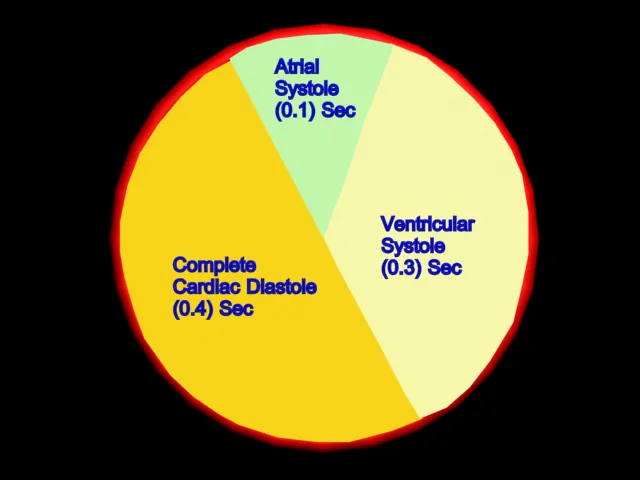
60 to 80. Taking 74 as an example each cycles lasts about 0.8 of a second and
consists of:
1) Atrial systole-
contraction of the atria.
2) Ventricular
systole- Contarction of the ventricles.
3)Complete cardiac
diastole- relaxation of the atria and ventricles
The superior vena
cava and the inferior vena cava transport deoxygenated blood into the right
atrium at the same time as the 4 pulmonary veins bring oxygenated blood into
the left atrium.The atrioventricular valves are open and blood flows passively
through to the ventricles.The SA node triggers a wave of contraction that
spreads over the myocardium of both atria,emptying the atria and completing
ventricular filling.When the electrical impulse reaches the AV node it is
slowed down,delaying
atrioventricular transmission.
This delay means that the mechanical result of the atrial stimulation,atrial contarction,lags behind the electrical activity by a fraction of a second.This allows the atria to finish emptying into the ventricles before the ventricles begin to contract.After this brief delay,the AV node triggers its own electrical impulse, which quickly spreads to the ventricular muscle via AV bundle,The bundle branches and Purkinje fibres.
This result in a wave of contraction which sweeps upwards from the apex of the heart and across the walls of both ventricles pumping the blood into the pulmonary artery and the aorta(Ventricular systole 0.3 sec).The high pressure generated during ventricular contraction is greater than that in the aorta and forces the atrioventricular valves to close preventing backflow of blood into the atria.
After contraction of
the ventricles there is complete cardiac diastole, a period of 0.4
secs,When atria and ventricles are relaxed.During this time the myocardium
recovers in preparation for the next heartbeat,and the atria refill in
preparation for the next cycle.
3D Animation Showing Blood Flow within Heart.
FLOW OF
BLOOD THROUGH THE HEART:
The two largest veins
of the body,the superior and inferior vena cava, empty their contents into
the right atrium.The blood passes via the right atrioventricular valve into the
right ventricle,and from there it is pumped into the pulmonary artery or trunk
(The only artery in the body which carries deoxygenated blood). The
opening of the pulmonary artery is
guarded by the pulmonary valve, formed by three semilunar cusps.The
valve prevents the backflow of blood into the backflow of blood
into the right ventricle when the ventricular muscle relaxes. After leaving the
heart the pulmonary artery divides into left nad right pulmonary arteries,
which carry the venous blood to the lungs where exchange of gases takes place;
carbon dioxide is excreted and oxygen is absorbed.
 |
| Heart Lung Tissue Circulation of Human Body |
Two pulmonary veins
from each lung carry oxygenated blood back to the left atrium.Blood then passes
through the left atrioventricular valve into the left ventricle, and from
there it is pumped into the aorta,the first artery of the general circulation.The
opening of the aorta is guarded by the aortic valve, formed by three semilunar
cusps.
From this sequence of
events it can be seen that the blood passes from the right to the left side of
the heart via the lungs or pulmonary circulation . However, it should be noted
that both atria contract at the same time and this is followed by the
simultaneous contraction of both ventricles.
The muscle layer of
the walls of the atria is thinner than that of the ventricles.This is
consistent with the amount of work they do. The atria usually assisted by
gavity,propel the blood to the lungs and round the whole body. The pulmonary
trunk leaves the heart from the upper part of the right ventricle,and the aorta
leaves from the upper part of the left ventricle.
 |
| SA Node and AV Node of Human Heart |
SA
NODE(SINOATRIAL NODE) AND AV NODE (ATRIOVENTRICULAR NODE):
The heart has an
intrinsic system whereby the cardiac muscle is automatically stimulated to contract
without the need of external stimulation.The property is called autorhythmicity.However,the
intrinsic system can be stimulated or depressed by nerve impulses initiated in
the brain and by circulating chemicals, including hormones.
3D Animation showing SA and AV Node of Heart.
3D Animation showing SA and AV Node of Heart.
Small group of
specialised neuromuscular cells in the myocardium initiate and conduct
impuses,causing coordinated and synchronised contraction of the heart muscle.
Sinoatreal
node(SA node):
This small mass of
specialised cells lies in the wall of the right atrium near the opening of the
superior vena cava.The SA node is pacemaker of the heart because it
normally initiates impulses more rapidly than other groups of neuromuscular
cells.Firing of the SA node causes atrial contraction.
Atrioventricular
node(AV node):
This small mass of
neuro muscular tissue is situated in the wall of the atrial septum near the
atrioventricular valves.Normally,the AV node conducts impulses that arrive via
the atria and that originated from the SA node.There is a delay here; the
electrical signal takes 0.1 of a second to pass through into the
ventricles.This allows the atria to finish contracting before the ventricles
start.
Atrioventricular
bundle(Bundle of His):
This is a mass of
specialised fibres that originate from the AV node. The AV bundle crosses the
fibrous ring that separates atria and ventricles then,at the upper end of the
ventricular septum,it divides into right and left bundle branches.Within the
ventricular myocardium the branches bundle, bundle branches and purkinje fibres
convey electrical impulses from the AV node to the apex of the myocardium where
the wave of ventricular contraction begins, then sweeps upwards and outwards pumping blood into
the pulmonary artery and aorta.
 |
| Blood Pressure Measuring Device |
BLOOD
PRESSURE:
This is
the force or pressure that the blood exerts on the walls of blood
vessels.Keeping blood pressure within normal limits is very important .If it
is high blood vessel can be damaged,causing clots or bleeding from sites of
blood vessel rupture.If it is falls to low ,then blood flow through tissue beds
may be inadequate.This is particularly dangerous for such essential organs as
the heart brain or kidney.
Blood
pressure varies according to the time of day, the posture,gender and age of the
individual.During bed rest at night the blood pressure tends to be lower.It
increases with age and is usually higher in women than in men. Atrial blood
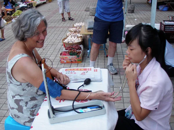
pressure is measured with a sphygmomanometer and is usually expressed with the systolic
pressure written above the diastolic pressure:
120 16
BP
=-------- mm Hg or BP = ----- kPa.
80
11
Cardiac Peripheral
Electric
Charges in the Heart:
As the
body fluid and tissues are good conductors of electricity, the electrical
activity within the heart can be detected by attaching electrodes to the
surface of the body. The Pattern of electrical activity may be displayed on an
oscilloscope screen or traced on paper. The apparatus used is an
electrocardiograph and the tracing is an electrocardiogram (ECG).
 |
| Electrocardiograph machine and ElectrocardiogramTracing |
The
normal ECG tracing show five waves which by convention, have been named P,Q,R,S
and T. The P wave arises when the impulse from the SA node sweeps over the
atria. The QRS complex represents the very rapid spread of the impulse from the
AV node through the AV bundle and the Purkinje fibers and the electrical
activity of the ventricular muscle.


 Online Movies
The KO Shop Australia Pty Ltd began its journey in 2014, as a boutique gift store. Since inception, The KO Shop directly imported and retailed high quality leather goods, hand crafted silver jewellery, singing bowls, wooden toys, clothing, handicraft works etc, from India, Nepal & Fiji. Over years, the range of imported goods expanded, and The KO Shop ventured into distribution
ReplyDeletesatya backflow cones and dhoop cones